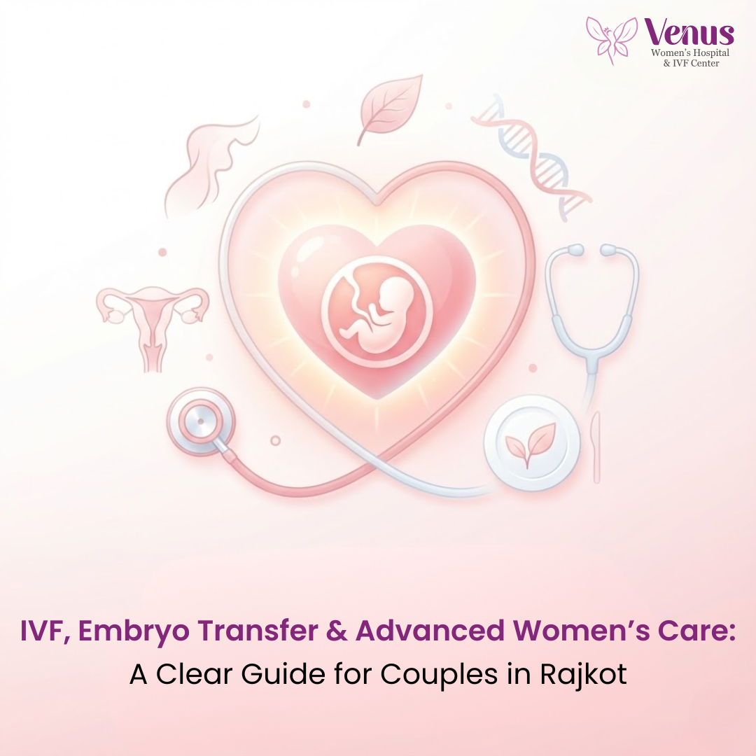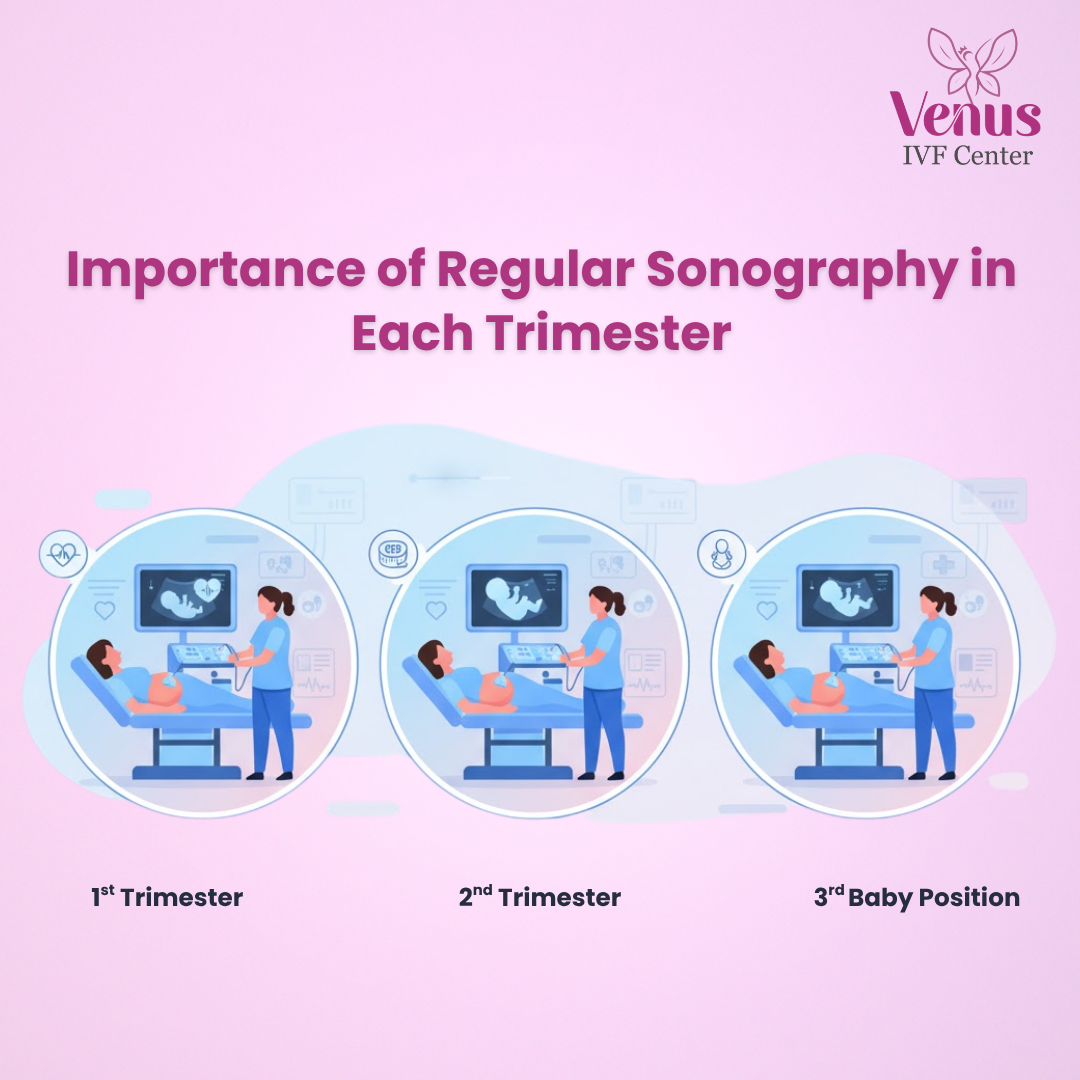
Pregnancy is a beautiful journey, but it also comes with many questions and uncertainties. One of the most important tools to ensure a healthy pregnancy is regular sonography. If you are expecting in Rajkot, understanding the significance of sonography at every stage helps you and your doctor monitor your baby’s growth and detect potential complications early.
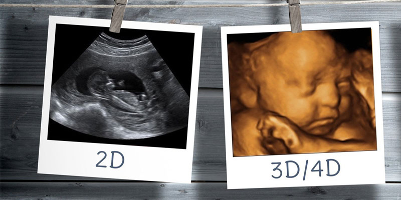
Prenatal sonography, commonly called an ultrasound scan, uses high-frequency sound waves to create images of the baby inside the womb. These scans are non-invasive, safe, and essential for assessing fetal health, growth, and development. At Venus Women’s Hospital in Rajkot, we offer advanced 2D, 3D, and 4D ultrasounds for expecting mothers, ensuring accurate and detailed information.
Regular sonography is not just about seeing your baby—it’s a medical necessity. Here’s why:
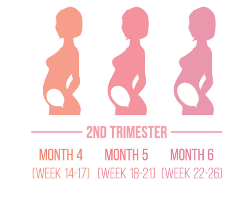
Monitoring Fetal Growth: Each trimester, your baby grows rapidly. Ultrasounds track weight, length, and organ development.
Purpose:
Recommended Scans:
Dating Scan: Confirms how far along you are and establishes an expected due date.
Tip for Mothers in Rajkot: Book your first sonography with a qualified gynecologist or pregnancy specialist to ensure accurate readings and expert guidance.
Purpose:
Recommended Scans:
Anomaly Scan (18–22 weeks): Provides detailed images of the fetal organs, spine, and limbs.
Local Insight: At Venus Women’s Hospital, our 3D and 4D ultrasound services in Rajkot allow expecting parents to view detailed, lifelike images of their baby, making the experience both informative and emotional.
Purpose:
Recommended Scans:
Expert Tip: Regular third-trimester scans help pregnancy doctors in Rajkot prepare for a safe and timely delivery, reducing risks for both mother and baby.
2D Ultrasound: Traditional, black-and-white images, excellent for basic assessment.
At Venus Women’s Hospital, all these technologies are available to provide comprehensive prenatal care.
Hydrate Well: A full bladder can improve image clarity, especially in early scans.
Choosing a trusted hospital for sonography is crucial. At Venus Women’s Hospital in Rajkot, we provide:
Experienced obstetricians and gynecologists
Our team ensures you receive accurate assessments and compassionate support throughout your pregnancy.
Regular sonography in each trimester is essential for a healthy pregnancy. It helps detect complications early, monitors fetal growth, and provides peace of mind for expecting mothers. For families in Rajkot, consulting a reliable pregnancy doctor and scheduling timely scans is the key to a safe and joyful pregnancy journey.
Book your next prenatal sonography session at Venus Women’s Hospital, Rajkot, and experience advanced, expert care for every stage of your pregnancy.
.jpg)

.jpg)


.png)

.jpg)


.jpg)

.png)
.png)


.png)


.jpg)


.png)
.png)

.png)
.png)