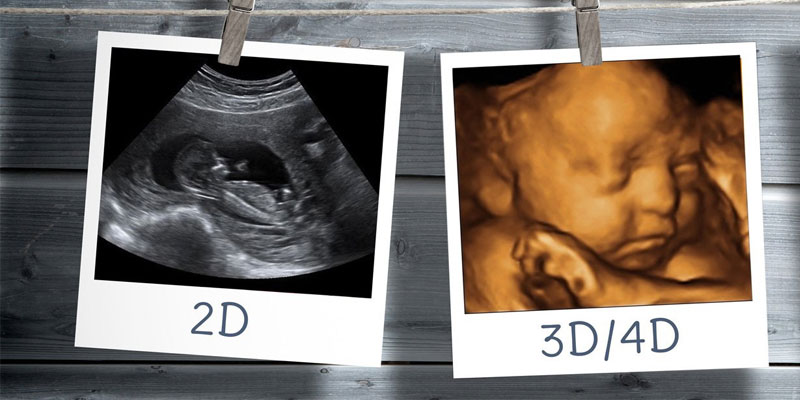
The journey of pregnancy is filled with moments of joy, anticipation, and wonder. Among these, the first glimpse of your baby through an ultrasound scan is perhaps the most thrilling. Traditionally, 2D sonography has been the norm for prenatal scans, offering black-and-white images that gave expectant parents a sneak peek of their developing baby. However, with advancements in medical imaging technology, 3D and 4D ultrasounds have transformed the way we view prenatal life, bringing your baby's first images to life with stunning clarity and detail.
To appreciate the magic of 3D and 4D ultrasounds, it’s essential to understand what these terms mean. A 3D (three-dimensional) ultrasound compiles multiple two-dimensional images taken at various angles to create a static three-dimensional image of the fetus. This technology allows parents to see their baby’s features more realistically, offering views of the face, limbs, and organs that were not possible with traditional 2D sonography.
The 4D ultrasound, meanwhile, adds a dynamic element - time. It captures moving 3D images, enabling you to see real-time activities of your baby, such as smiling, yawning, and kicking. This technology provides an incredibly intimate view of the baby's in-utero life, making the experience even more special for expectant parents.
The evolution from 2D to 3D and 4D ultrasounds marks a significant leap in medical imaging. While 2D sonography remains a critical tool for assessing fetal health and development, 3D and 4D technologies have enhanced the emotional and bonding experience of parents with their unborn child. The journey from seeing a flat, often indistinct image to a vivid, lifelike portrayal of the fetus is nothing short of magical.
The journey began with 2D (two-dimensional) ultrasound technology. This traditional form of sonography, still widely used today, provides flat, black-and-white images of the fetus. It uses sound waves that bounce off internal structures to create cross-sectional images of the uterus, placenta, and fetus. These images are invaluable for assessing the baby's growth, position, and basic anatomy, as well as the mother's reproductive organs. However, 2D ultrasounds have their limitations in terms of the depth and detail of the image.
The introduction of 3D (three-dimensional) ultrasound marked a significant leap forward. This technology uses sophisticated software to process multiple two-dimensional images taken from different angles, creating a three-dimensional image of the fetus. These images offer a more realistic view of the baby’s facial features and anatomy, providing a clearer picture for both parents and healthcare providers. 3D sonography has become particularly useful in diagnosing certain fetal anomalies that may not be as evident in 2D images, such as cleft lip or spinal abnormalities.
The evolution continued with the advent of 4D (four-dimensional) ultrasound, which adds the element of time to 3D images, essentially creating a live video of the fetus in the womb. This allows parents and medical professionals to see movements and behaviors in real-time, such as smiling, yawning, or sucking a thumb. This dynamic aspect of 4D sonography provides a new depth of prenatal bonding and also aids in the assessment of fetal behavior and development.
While 3D and 4D ultrasounds have provided new insights into fetal development, it's important to note that 2D ultrasounds remain the standard in prenatal care due to their proven effectiveness in medical diagnostics. Generally Obstetricians and Gynecologists and other health authorities consider 3D and 4D ultrasounds to be safe when used appropriately by trained professionals, primarily for medical purposes rather than just keepsakes.
3D and 4D ultrasounds are not just about getting a beautiful image of the baby; they have substantial clinical applications too. These advanced sonography techniques can provide better insights into fetal anatomy, helping healthcare professionals in diagnosing potential issues more accurately. Conditions such as cleft lip, spinal cord issues, and heart defects can be more easily identified with 3D/4D imaging.
Moreover, these ultrasounds can be instrumental in planning medical or surgical interventions that may be required after birth. They also help in better understanding the position of the placenta and umbilical cord, which can be crucial for delivery planning.
When it comes to the safety of 3D and 4D ultrasounds, the general consensus among medical professionals is that they are safe when used appropriately. However, it’s important to note that these should be performed by qualified healthcare professionals.
No Proven Risks but Caution Advised: There are no known risks specifically associated with 3D and 4D ultrasounds in pregnancy. However, like all medical procedures, they should be performed with caution. The American College of Obstetricians and Gynecologists (ACOG) and the Food and Drug Administration (FDA) advise that ultrasounds should be performed only when medically necessary.
Heat and Bubble Formation: Ultrasounds work by sending sound waves into the body, which can cause slight heating and, in some cases, the formation of small bubbles in tissues.
Qualified Medical Professionals: All ultrasounds, including 3D and 4D, should be performed by qualified healthcare professionals. They are trained to use the equipment safely and to interpret the images accurately, ensuring both the safety of the procedure and the reliability of the results.
Avoid Keepsake Ultrasounds: The FDA strongly discourages the use of 3D and 4D ultrasounds for creating keepsake videos or photos. This is because non-medical settings might not follow the same stringent safety protocols as medical facilities, and staff may not be adequately trained to operate ultrasound machines safely.
The emotional impact of 3D and 4D ultrasounds on expectant parents cannot be overstated. Seeing detailed images or videos of the baby in the womb often deepens the bond between the parents and the child. It makes the pregnancy more real, especially for partners who might not be experiencing the physical changes associated with pregnancy.
If you’re considering 3D or 4D ultrasound hospital, it’s essential to choose the right facility. Ensure that the sonography center is reputable, equipped with the latest technology, and staffed by experienced professionals. It’s always a good idea to consult with your healthcare provider before scheduling an elective 3D or 4D ultrasound.
The advent of 3D and 4D ultrasound technology has revolutionized prenatal imaging, offering expectant parents a magical and intimate view of their developing baby. While the primary aim of these scans should always be to ensure the health and well-being of the fetus, the joy and wonder they bring to parents are immeasurable. As we continue to advance in medical technology, the ability to connect with and understand life within the womb will only deepen, strengthening the bond between parents and their baby before they even enter the world.
.jpg)

.jpg)


.png)

.jpg)


.jpg)


.png)
.png)


.png)


.jpg)


.png)


.png)

.png)
.png)